Myopia, also known as nearsightedness, is a common eye condition that affects millions of people worldwide. It occurs when the eyeball is too long or the cornea is too curved, causing light to focus in front of the retina instead of on it. As a result, objects in the distance appear blurry, while those up close are clear.

Myopia Symptoms

Symptoms of myopia can include squinting, headaches, eye strain, and difficulty seeing objects in the distance. Children with myopia may also have trouble seeing the board in school or playing sports that require good distance vision.
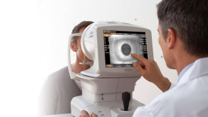
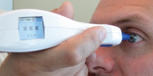
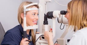
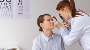
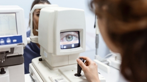
Myopia vs Normal Diagram

Diagnosing Myopia

Diagnosing myopia typically involves a comprehensive eye exam, which may include a visual acuity test, refraction test, and measurement of the shape of the cornea and the length of the eyeball. Early detection is important, as untreated myopia can lead to serious long-term complications.

Risk Factors Associated with Myopia

Risk factors for myopia include genetics, age, and certain environmental factors. Children with one or both parents who have myopia are more likely to develop the condition themselves. Spending a lot of time doing near work, such as reading or using a computer, may also increase the risk of myopia.

Long Term Implications of Myopia

Long-term implications of myopia can include an increased risk of cataracts, glaucoma, and retinal detachment. Additionally, high levels of myopia can lead to a condition called myopic maculopathy, which can cause permanent vision loss.

Myopia Prevention

Preventing myopia is difficult, as many risk factors are beyond our control. However, there is some evidence to suggest that spending more time outdoors and limiting near work may help reduce the risk of myopia in children.

Myopia Treatment Options

Treatment options for myopia include corrective lenses, such as glasses or contact lenses, and refractive surgery. Glasses or contacts work by bending light to focus it correctly on the retina, while refractive surgery reshapes the cornea to achieve the same result. Additionally, orthokeratology involves wearing special contact lenses that reshape the cornea overnight to temporarily improve vision during the day.

In conclusion, myopia is a common eye condition that can have serious long-term implications if left untreated. Early detection and treatment are important to prevent complications and preserve vision. While preventing myopia is difficult, certain lifestyle changes may help reduce the risk in children. If you are experiencing symptoms of myopia, it is important to schedule an eye exam with a qualified eye care professional.

LASIK (Laser-Assisted In Situ Keratomileusis) is a surgical procedure that reshapes the cornea of the eye to correct vision problems such as nearsightedness, farsightedness, and astigmatism. LASIK surgery has been a popular option for people seeking to improve their vision without the use of glasses or contact lenses. In this article, we will discuss the different eye conditions that benefit from LASIK eye surgery.

1. Nearsightedness (Myopia)

Nearsightedness, also known as myopia, is a common vision problem that affects millions of people worldwide. In this condition, the person can see nearby objects clearly, but distant objects appear blurry. LASIK surgery corrects nearsightedness by reshaping the cornea to focus the light more accurately on the retina, resulting in clearer vision.

2. Farsightedness (Hyperopia)

Farsightedness, also known as hyperopia, is a condition in which the person can see distant objects clearly, but nearby objects appear blurry. This condition occurs when the eyeball is too short, or the cornea is too flat, resulting in light focusing behind the retina instead of on it. LASIK surgery can correct farsightedness by reshaping the cornea to focus the light more accurately on the retina, resulting in clearer vision.

3. Astigmatism

Astigmatism is a common vision problem in which the cornea is irregularly shaped, resulting in distorted or blurred vision. This condition can cause headaches, eye strain, and difficulty seeing at night. LASIK surgery can correct astigmatism by reshaping the cornea to create a more regular shape, resulting in clearer vision.

4. Presbyopia

Presbyopia is an age-related condition in which the eye’s natural lens becomes less flexible, making it difficult to focus on close-up objects. This condition typically affects people over the age of 40 and can cause difficulty with reading, using a computer, or doing close-up work. LASIK surgery can correct presbyopia by reshaping the cornea to create a multifocal surface, allowing the person to see both near and distant objects clearly.

5. Keratoconus

Keratoconus is a progressive eye disease that causes the cornea to thin and bulge into a cone shape, resulting in distorted vision. In some cases, this condition can be treated with LASIK surgery to reshape the cornea and improve vision. However, not all patients with keratoconus are suitable candidates for LASIK surgery, and other treatments may be more appropriate.

In conclusion, LASIK eye surgery can be a safe and effective way to correct various vision problems, including nearsightedness, farsightedness, astigmatism, presbyopia, and, in some cases, keratoconus. However, it is important to consult with an experienced ophthalmologist to determine if LASIK surgery is the right option for you based on your individual eye condition and other factors.

An autorefractor is a medical instrument used to measure the refractive error of the eye, which is the degree of nearsightedness, farsightedness, or astigmatism that affects a person’s vision. It is commonly used by optometrists and ophthalmologists to obtain an objective measurement of a patient’s refractive error, which can be used to determine the appropriate prescription for eyeglasses or contact lenses.

The autorefractor uses advanced technology to quickly and accurately measure the way light is refracted, or bent, as it passes through the eye’s optical system. The instrument works by projecting a beam of light into the eye and measuring how the light is reflected back. The autorefractor uses a series of lenses and mirrors to calculate the degree of refractive error based on the pattern of light that is reflected back.

The autorefractor is a non-invasive and painless procedure that typically takes only a few minutes to complete. The patient simply sits in front of the instrument and focuses on a target while the examiner positions the autorefractor to align with the eye. The instrument emits a series of flashes of light, and the patient may be asked to blink or look in different directions to obtain a more accurate measurement.

Uses for a Autorefractor

The autorefractor can provide an objective measurement of the refractive error that is not influenced by the patient’s responses or subjective factors, such as fatigue, anxiety, or inexperience with vision testing. It can also provide a more precise measurement than traditional manual methods, such as the use of a phoropter, which requires the patient to make subjective judgments about the clarity and sharpness of different lenses.

The autorefractor is especially useful in diagnosing and correcting refractive errors in children and adults who may have difficulty communicating their vision problems or may have cognitive or developmental challenges that make traditional vision testing more difficult. It can also be used to monitor changes in the refractive error over time and adjust the prescription as needed.

In summary, an autorefractor is a valuable tool for measuring the refractive error of the eye and obtaining an objective measurement of the patient’s vision. It is a non-invasive and painless procedure that can provide a more precise measurement than traditional manual methods and is especially useful in diagnosing and correcting vision problems in children and adults. If you are experiencing vision problems or are due for a routine eye exam, your optometrist or ophthalmologist may use an autorefractor to assess your refractive error and help you achieve clear, comfortable vision.

An ophthalmoscope is a medical instrument used by eye doctors, such as optometrists and ophthalmologists, to examine the internal structures of the eye, including the retina, optic nerve, and blood vessels. It allows the practitioner to view the inside of the eye and detect any abnormalities or signs of disease, such as macular degeneration, glaucoma, or diabetic retinopathy. The ophthalmoscope can also be used to measure the refractive error and assess the need for corrective lenses.

The ophthalmoscope consists of a light source, a lens system, and a viewing aperture or eyepiece. The light source is usually a halogen or LED bulb that emits a bright, focused beam of light. The lens system includes a series of lenses and filters that focus the light and adjust its intensity and color. The viewing aperture or eyepiece allows the examiner to see through the instrument and into the eye.

To use the ophthalmoscope, the patient sits in a darkened room and focuses on a distant object while the examiner approaches with the instrument. The examiner holds the ophthalmoscope close to the patient’s eye and shines the light into the pupil, which dilates in response to the bright light. The examiner then adjusts the lenses and filters to focus the light on the retina and visualize its structures.

Uses for a Ophthalmoscope

The ophthalmoscope can reveal many important details about the eye, including the condition of the optic nerve, the presence of any retinal tears or holes, and the status of the blood vessels that supply the retina. The examiner can also observe the macula, which is the small area at the center of the retina that is responsible for sharp, detailed vision. Any abnormalities in the macula can indicate conditions such as macular degeneration or diabetic retinopathy.

In addition to diagnosing eye diseases, the ophthalmoscope can also be used to measure the refractive error of the eye and assess the need for corrective lenses. By adjusting the lenses and filters of the instrument, the examiner can determine the strength and type of lenses needed to correct myopia (nearsightedness), hyperopia (farsightedness), or astigmatism.

In summary, the ophthalmoscope is a valuable tool for examining the internal structures of the eye and detecting signs of disease or abnormalities. It allows eye doctors to diagnose and treat a wide range of conditions and provide customized care for each patient’s individual needs. If you are experiencing vision problems or have a family history of eye disease, it is important to schedule regular eye exams with a qualified eye doctor who can use an ophthalmoscope to assess your eye health and help you maintain clear, healthy vision.

A keratometer is an ophthalmic instrument used to measure the curvature of the cornea, which is the clear front surface of the eye that covers the iris and the pupil. It is also known as an ophthalmometer, a corneal topographer, or a manual keratometer. The device is commonly used by optometrists and ophthalmologists to determine the refractive error and the astigmatism of the eye, as well as to fit contact lenses and assess the candidacy for refractive surgery.

The keratometer consists of a pair of movable arms or spindles that are attached to a central axis, a light source, and a scale or dial. The patient sits in front of the instrument and focuses on a target while the examiner positions the instrument to align with the cornea. The light source projects a pattern of concentric circles or rings onto the cornea, which reflects back through the instrument and is viewed through a magnifying eyepiece.

The keratometer measures the radius of curvature of the cornea in two meridians, or directions, usually the horizontal and the vertical, but sometimes other oblique angles as well. The curvature is expressed in diopters, which is a unit of measurement of the refractive power of a lens. A diopter is defined as the reciprocal of the focal length of a lens in meters, so a keratometer measures the curvature of the cornea as if it were a lens.

Uses for a Keratometer

The keratometer is especially useful in diagnosing and correcting astigmatism, which is a common refractive error that occurs when the cornea is not perfectly spherical, but has a more oblong or elliptical shape. Astigmatism can cause blurred or distorted vision, especially at certain distances or angles. The keratometer can measure the degree and orientation of the astigmatism, which can be corrected with cylindrical lenses or toric contact lenses.

The keratometer can also be used to assess the corneal topography, which is the three-dimensional shape and curvature of the cornea. Corneal topography is important for detecting and monitoring corneal diseases, such as keratoconus, which is a progressive thinning and bulging of the cornea that can cause severe visual impairment. The keratometer can help in the early detection and treatment of keratoconus by mapping the corneal shape and thickness.

In summary, a keratometer is a valuable tool for measuring the curvature of the cornea and diagnosing refractive errors, astigmatism, and corneal diseases. It is a non-invasive and relatively simple instrument that can provide important information about the eye’s optical properties and guide the selection of appropriate treatments and corrective lenses. If you are experiencing vision problems or are considering contact lenses or refractive surgery, your optometrist or ophthalmologist may use a keratometer to assess your corneal curvature and help you achieve optimal vision.

The pachymeter is a diagnostic tool used in ophthalmology to measure the thickness of the cornea, the clear outer layer of the eye. This simple, yet powerful device has become an essential tool in the diagnosis and treatment of a variety of eye conditions.

The cornea plays a crucial role in vision by refracting light as it enters the eye. Any irregularities in its shape or thickness can lead to blurry or distorted vision. The pachymeter measures the thickness of the cornea, providing valuable information to ophthalmologists in diagnosing and managing corneal disorders.

The pachymeter is a handheld device that uses ultrasound waves to measure the thickness of the cornea. The device emits a high-frequency sound wave that passes through the cornea and reflects back to the pachymeter. By analyzing the time it takes for the sound wave to return, the device can calculate the thickness of the cornea.

Uses for a Pachymeter

One of the most common conditions diagnosed using the pachymeter is glaucoma. Glaucoma is a condition where the pressure inside the eye increases, causing damage to the optic nerve and leading to vision loss. The pachymeter is used to measure the thickness of the cornea, which can affect the accuracy of intraocular pressure measurements. Thinner corneas can give falsely low pressure readings, while thicker corneas can give falsely high pressure readings. By taking the corneal thickness into account, ophthalmologists can more accurately diagnose and manage glaucoma.

The pachymeter is also used to monitor the progression of corneal disorders such as keratoconus. Keratoconus is a condition where the cornea becomes thinner and more conical in shape, leading to distorted vision. The pachymeter is used to measure the thickness of the cornea at various points, allowing ophthalmologists to monitor the progression of the disease and determine the appropriate treatment.

In addition to its diagnostic and monitoring capabilities, the pachymeter is also used to guide certain treatments. For example, during corneal transplant surgery, the pachymeter is used to measure the thickness of the donor cornea and ensure that it is a good match for the recipient’s cornea.

In conclusion, the pachymeter is a valuable tool in the field of ophthalmology. By providing accurate measurements of corneal thickness, it allows ophthalmologists to more accurately diagnose and manage a variety of eye conditions. With its ability to monitor the progression of diseases and guide treatments, the pachymeter has become an essential tool in the care of the eyes.

The wavefront analyzer is a revolutionary tool in the field of ophthalmology. It is an imaging device that provides detailed information about the optical system of the eye, allowing ophthalmologists to diagnose and treat a variety of visual disorders. By analyzing the wavefront data, ophthalmologists can create customized treatment plans that improve the patient’s vision and quality of life.

The wavefront analyzer measures the way that light travels through the eye, providing a detailed map of the optical system. The device works by sending a beam of light into the eye and analyzing the pattern of light waves that are reflected back. This data is then used to create a three-dimensional map of the eye’s optical system, including the cornea, lens, and retina.

What is the Wavefront Analyser used for?

One of the primary uses of the wavefront analyzer is to diagnose and treat refractive errors. Refractive errors occur when the eye’s optical system is not perfectly shaped, leading to blurry or distorted vision. Common refractive errors include myopia (nearsightedness), hyperopia (farsightedness), and astigmatism. By analyzing the wavefront data, ophthalmologists can create customized treatment plans that correct these refractive errors, improving the patient’s vision.

Another use of the wavefront analyzer is to diagnose and treat higher-order aberrations. Higher-order aberrations are more complex visual problems that cannot be corrected with traditional glasses or contact lenses. These aberrations can include halos, starbursts, and double vision. By analyzing the wavefront data, ophthalmologists can create customized treatment plans improving the patient’s visual quality.

The wavefront analyzer can also be used to monitor the progress of certain treatments, such as LASIK surgery. LASIK is a popular refractive surgery that reshapes the cornea to correct refractive errors. By analyzing the wavefront data before and after surgery, ophthalmologists can track the progress of the healing process and ensure that the patient’s vision is improving as expected.

Capabilities of the Wavefront Analyser

In addition to its diagnostic and treatment capabilities, the wavefront analyzer has also been used in research to better understand the human eye. By analyzing the wavefront data researchers can gain insights into how the eye works and how it can be improved.

In conclusion, the wavefront analyzer is a powerful tool in the field of ophthalmology. By providing detailed information about the eye, it allows ophthalmologists to diagnose and treat a variety of visual disorders. With its ability to monitor the progress of treatments, the wavefront analyzer has revolutionized the way we approach vision care.

The corneal topographer is a vital diagnostic tool in modern ophthalmology. It is an imaging device that provides detailed information about the shape and curvature of the cornea, the clear outer layer of the eye. By analyzing the corneal topography, ophthalmologists can diagnose a variety of conditions, including astigmatism, keratoconus, and other corneal irregularities.

The cornea plays a crucial role in vision by refracting light as it enters the eye. Any irregularities in its shape can lead to blurry or distorted vision, and in some cases, even complete vision loss. The corneal topographer provides detailed measurements of the cornea’s curvature, which helps ophthalmologists diagnose and treat corneal disorders more effectively.

Corneal topography is a non-invasive procedure that involves placing a special camera in front of the eye. The camera captures multiple images of the cornea from different angles, and sophisticated software analyzes the data to create a detailed map of the corneal surface. The resulting map shows the curvature, thickness, and shape of the cornea, allowing ophthalmologists to diagnose corneal disorders accurately.

Corneal topography and Astigmatism

One of the most common conditions diagnosed using corneal topography is astigmatism. Astigmatism is a condition where the cornea is not perfectly round, leading to distorted or blurry vision. By analyzing the corneal topography, ophthalmologists can determine the severity and type of astigmatism, which helps them choose the most appropriate treatment.

Corneal topography and Keratoconus

Keratoconus is a condition that can be diagnosed using corneal topography. Keratoconus is a progressive condition where the cornea becomes thinner and more conical in shape, leading to distorted vision. Corneal topography can help diagnose keratoconus in its early stages, allowing for more effective treatment and management.

Corneal topography can be used to monitor the progress of certain treatments. After corneal transplant surgery, ophthalmologists use corneal topography to track the healing process and ensure that the new cornea is taking the proper shape.

The corneal topographer is an essential diagnostic tool in modern ophthalmology. By providing detailed information about the shape and curvature of the cornea, it helps ophthalmologists diagnose a variety of conditions, including astigmatism, keratoconus, and other corneal irregularities. With its non-invasive nature and ability to monitor the progress of treatments, corneal topography has revolutionized the diagnosis and treatment of corneal disorders.

In this article we detail the most popular LASIK diagnostic equipment and devices. We explore what they are used for and examine their specific functions in determining if a patient is a suitable candidate for LASIK surgery. Some common LASIK diagnostic devices include:

Corneal Topographer

A corneal topographer creates a detailed map of the surface of the cornea. It measures the curvature of the cornea, identifies irregularities and helps the surgeon plan the precise location and depth of the laser treatment.

The corneal topographer is a vital diagnostic tool in modern ophthalmology. It is an imaging device that provides detailed information about the shape and curvature of the cornea. By analyzing the corneal topography, ophthalmologists can diagnose a variety of conditions, including astigmatism, keratoconus, and other corneal irregularities.

Wavefront Analyzer

A wavefront analyser is a device that measures the unique optical characteristics of the patient’s eye, including how light travels through the eye and how it’s focused on the retina. The information gathered is used to create a customized LASIK treatment plan.

The device works by sending a beam of light into the eye and analyzing the pattern of light waves that are reflected back. This data is then used to create a three-dimensional map of the eye’s optical system, including the cornea, lens, and retina.

Pachymeter

This device measures the thickness of the cornea. A thin cornea can increase the risk of complications during surgery, so this measurement is important to determine whether the patient is a good candidate for LASIK.

The pachymeter is also used to monitor the progression of corneal disorders such as keratoconus. Keratoconus is a condition where the cornea becomes thinner and more conical in shape, leading to distorted vision.

Keratometer

A keratometer is an ophthalmic instrument used to measure the curvature of the cornea, which is the clear front surface of the eye that covers the iris and the pupil. It is also known as an ophthalmometer, a corneal topographer, or a manual keratometer.

The keratometer consists of a pair of movable arms or spindles that are attached to a central axis, a light source, and a scale or dial. The patient sits in front of the instrument and focuses on a target. The examiner positions the instrument to align with the cornea. The light source projects a pattern of concentric circles onto the cornea, which reflects back through the instrument and is viewed through a magnifying eyepiece.

Ophthalmoscope

This device allows the surgeon to examine the interior of the eye, including the retina and optic nerve. It’s important to check for any abnormalities that could impact the success of the LASIK surgery.

The ophthalmoscope consists of a light source, a lens system, and a viewing aperture or eyepiece. The light source is usually a halogen or LED bulb that emits a bright, focused beam of light. The lens system includes a series of lenses and filters that focus the light and adjust its intensity and color. The viewing aperture or eyepiece allows the examiner to see through the instrument and into the eye.

Autorefractor

This device measures the refractive error of the eye, which determines the patient’s prescription for glasses or contact lenses. Measurements can be used to guide the lasik procedure.

The autorefractor uses advanced technology to quickly and accurately measure the way light is refracted, or bent, as it passes through the eye. The instrument works by projecting a beam of light into the eye and measuring how the light is reflected back. The autorefractor uses a series of lenses and mirrors to calculate the degree of refractive error based on the pattern of light that is reflected back.

LASIK eye surgery is a type of refractive surgery that aims to correct vision problems such as nearsightedness, farsightedness, and astigmatism. LASIK, which stands for Laser-Assisted in Situ Keratomileusis, is a popular elective procedure that has helped millions of people around the world improve their vision.

In this article, we’ll take a closer look at what LASIK eye surgery is, how it works, and who is a good candidate for the procedure. We’ll also discuss the benefits and risks of LASIK, as well as what to expect during the recovery period.

LASIK Eye Surgery

LASIK eye surgery is a type of refractive surgery that uses a laser to reshape the cornea, which is the clear front surface of the eye. By reshaping the cornea, LASIK can help to improve the way that light enters the eye, allowing it to focus properly on the retina at the back of the eye.

LASIK is a popular choice for people who suffer from vision problems such as nearsightedness, farsightedness, and astigmatism. Nearsightedness, also known as myopia, occurs when the eye is too long, causing light to focus in front of the retina. Farsightedness, or hyperopia, occurs when the eye is too short, causing light to focus behind the retina. Astigmatism is a condition in which the cornea is irregularly shaped, causing blurred vision.

How Does LASIK Eye Surgery Work?

LASIK eye surgery works by using a laser to create a thin flap in the cornea. The flap is then lifted, and a laser is used to reshape the cornea, allowing it to better focus light onto the retina. After the cornea has been reshaped, the flap is replaced, and the eye is allowed to heal.

There are several different types of lasers that can be used during LASIK eye surgery, including excimer lasers, femtosecond lasers, and wavefront-guided lasers. Excimer lasers use ultraviolet light to remove small amounts of tissue from the cornea, while femtosecond lasers use pulses of laser energy to create a precise incision in the cornea. Wavefront-guided lasers use advanced computer technology to create a custom treatment plan for each patient, taking into account the unique characteristics of their eyes.

Who is a Good Candidate for LASIK Eye Surgery?

Not everyone is a good candidate for LASIK eye surgery. The ideal candidate is someone who is over 18 years old and has stable vision. They should not have any underlying eye diseases or conditions, such as glaucoma or cataracts, that could affect the outcome of the procedure. They should also have a healthy cornea, with enough thickness to allow for the creation of a corneal flap.

Not everyone is a good candidate for LASIK eye surgery. The ideal candidate is someone who is over 18 years old and has stable vision. They should not have any underlying eye diseases or conditions, such as glaucoma or cataracts, that could affect the outcome of the procedure. They should also have a healthy cornea, with enough thickness to allow for the creation of a corneal flap.

In addition, the ideal candidate for LASIK should have a strong desire to reduce their dependence on glasses or contact lenses. While LASIK can help to improve vision, it is not a guarantee that a person will no longer need corrective lenses. Some people may still need to wear glasses or contacts after the procedure, albeit with a lower prescription.

It’s important to note that LASIK is not recommended for everyone. Women who are pregnant or nursing should not undergo LASIK, as hormonal changes can affect the stability of their vision. People with certain medical conditions, such as autoimmune diseases or diabetes, may also be advised against LASIK.

Benefits and Risks of LASIK Eye Surgery

The benefits of LASIK eye surgery are numerous. For many people, LASIK can provide a significant improvement in vision, allowing them to see more clearly without the need for glasses or contacts. The procedure is also relatively quick and painless, with most people experiencing only minor side effects.