In this article we detail the most popular LASIK diagnostic equipment and devices. We explore what they are used for and examine their specific functions in determining if a patient is a suitable candidate for LASIK surgery. Some common LASIK diagnostic devices include:
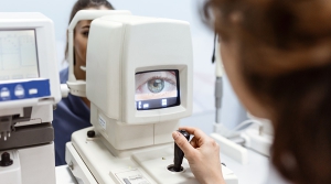
Corneal Topographer
A corneal topographer creates a detailed map of the surface of the cornea. It measures the curvature of the cornea, identifies irregularities and helps the surgeon plan the precise location and depth of the laser treatment.
The corneal topographer is a vital diagnostic tool in modern ophthalmology. It is an imaging device that provides detailed information about the shape and curvature of the cornea. By analyzing the corneal topography, ophthalmologists can diagnose a variety of conditions, including astigmatism, keratoconus, and other corneal irregularities.

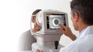
Wavefront Analyzer
A wavefront analyser is a device that measures the unique optical characteristics of the patient’s eye, including how light travels through the eye and how it’s focused on the retina. The information gathered is used to create a customized LASIK treatment plan.
The device works by sending a beam of light into the eye and analyzing the pattern of light waves that are reflected back. This data is then used to create a three-dimensional map of the eye’s optical system, including the cornea, lens, and retina.
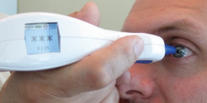
Pachymeter
This device measures the thickness of the cornea. A thin cornea can increase the risk of complications during surgery, so this measurement is important to determine whether the patient is a good candidate for LASIK.
The pachymeter is also used to monitor the progression of corneal disorders such as keratoconus. Keratoconus is a condition where the cornea becomes thinner and more conical in shape, leading to distorted vision.
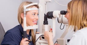
Keratometer
A keratometer is an ophthalmic instrument used to measure the curvature of the cornea, which is the clear front surface of the eye that covers the iris and the pupil. It is also known as an ophthalmometer, a corneal topographer, or a manual keratometer.
The keratometer consists of a pair of movable arms or spindles that are attached to a central axis, a light source, and a scale or dial. The patient sits in front of the instrument and focuses on a target. The examiner positions the instrument to align with the cornea. The light source projects a pattern of concentric circles onto the cornea, which reflects back through the instrument and is viewed through a magnifying eyepiece.
Ophthalmoscope
This device allows the surgeon to examine the interior of the eye, including the retina and optic nerve. It’s important to check for any abnormalities that could impact the success of the LASIK surgery.
The ophthalmoscope consists of a light source, a lens system, and a viewing aperture or eyepiece. The light source is usually a halogen or LED bulb that emits a bright, focused beam of light. The lens system includes a series of lenses and filters that focus the light and adjust its intensity and color. The viewing aperture or eyepiece allows the examiner to see through the instrument and into the eye.

Autorefractor
This device measures the refractive error of the eye, which determines the patient’s prescription for glasses or contact lenses. Measurements can be used to guide the lasik procedure.
The autorefractor uses advanced technology to quickly and accurately measure the way light is refracted, or bent, as it passes through the eye. The instrument works by projecting a beam of light into the eye and measuring how the light is reflected back. The autorefractor uses a series of lenses and mirrors to calculate the degree of refractive error based on the pattern of light that is reflected back.