An autorefractor is a medical instrument used to measure the refractive error of the eye, which is the degree of nearsightedness, farsightedness, or astigmatism that affects a person’s vision. It is commonly used by optometrists and ophthalmologists to obtain an objective measurement of a patient’s refractive error, which can be used to determine the appropriate prescription for eyeglasses or contact lenses.
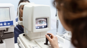
The autorefractor uses advanced technology to quickly and accurately measure the way light is refracted, or bent, as it passes through the eye’s optical system. The instrument works by projecting a beam of light into the eye and measuring how the light is reflected back. The autorefractor uses a series of lenses and mirrors to calculate the degree of refractive error based on the pattern of light that is reflected back.
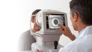
The autorefractor is a non-invasive and painless procedure that typically takes only a few minutes to complete. The patient simply sits in front of the instrument and focuses on a target while the examiner positions the autorefractor to align with the eye. The instrument emits a series of flashes of light, and the patient may be asked to blink or look in different directions to obtain a more accurate measurement.
Uses for a Autorefractor
The autorefractor can provide an objective measurement of the refractive error that is not influenced by the patient’s responses or subjective factors, such as fatigue, anxiety, or inexperience with vision testing. It can also provide a more precise measurement than traditional manual methods, such as the use of a phoropter, which requires the patient to make subjective judgments about the clarity and sharpness of different lenses.
The autorefractor is especially useful in diagnosing and correcting refractive errors in children and adults who may have difficulty communicating their vision problems or may have cognitive or developmental challenges that make traditional vision testing more difficult. It can also be used to monitor changes in the refractive error over time and adjust the prescription as needed.
In summary, an autorefractor is a valuable tool for measuring the refractive error of the eye and obtaining an objective measurement of the patient’s vision. It is a non-invasive and painless procedure that can provide a more precise measurement than traditional manual methods and is especially useful in diagnosing and correcting vision problems in children and adults. If you are experiencing vision problems or are due for a routine eye exam, your optometrist or ophthalmologist may use an autorefractor to assess your refractive error and help you achieve clear, comfortable vision.